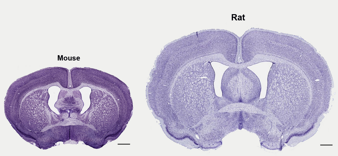
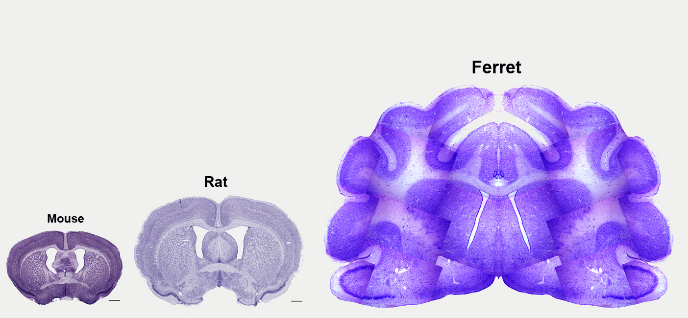
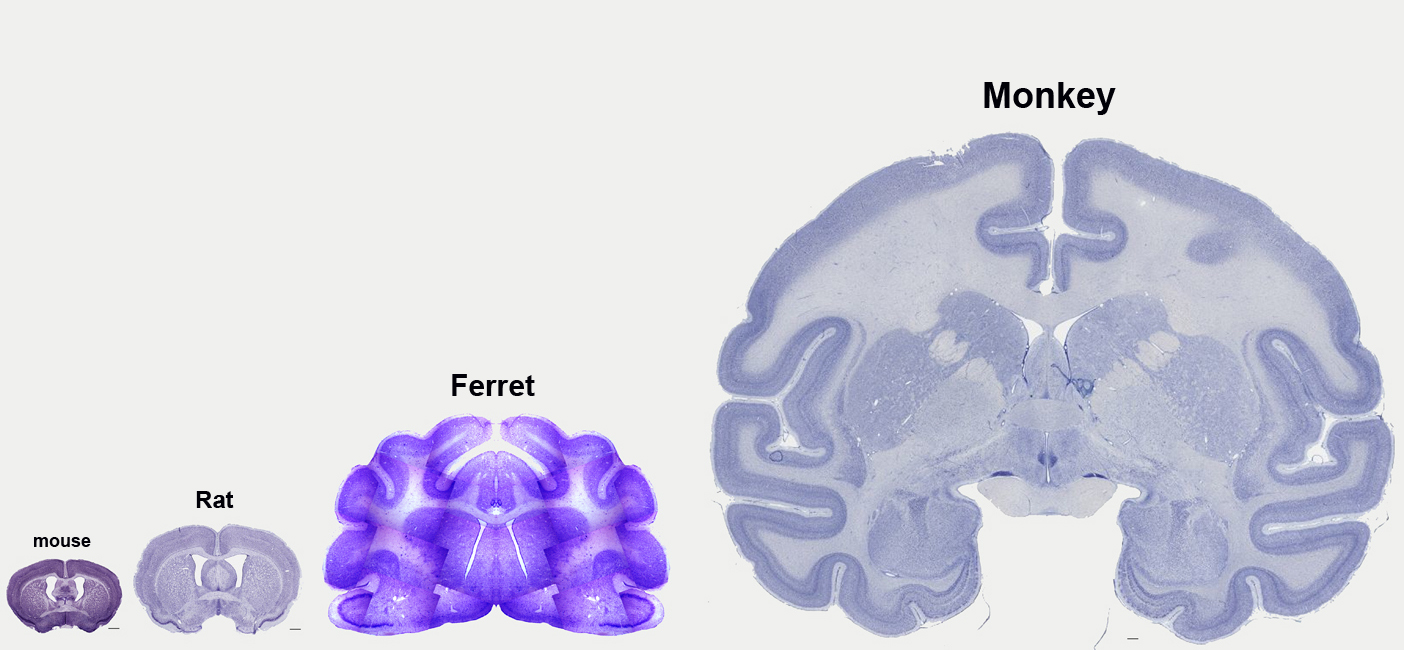
The models commonly used in researching brain function and development are substantially different. This is of course stating the obvious when considering mouse and human. But even mouse and rat are quite different. Shown below for comparison, are are Nissl stained sections of mature brain tissue from mouse, rat, ferret, rhesus monkey, and human.
Each section was taken from about the level of the anterior commissure, and in each image the sections are shown at the same scale. In the last image you can appreciate the obvious difference in size between human and mouse brain (and everything in between), but in the first image you will also note that the rat brain is significantly larger than the mouse brain. In fact, the rat cerebral cortex has about three times more neurons than the mouse cortex. Recent estimates indicate that there are ~10 million neurons in the mouse cerebral cortex (see Herculano-Houzel, Watson and Paxinos, Frontiers in Neuroanatomy, 2013), and ~30 million neurons in the rat cerebral cortex (see Mortera and Herculano-Houzel, Frontiers in Neuroanatomy, 2012). Something to be aware of…
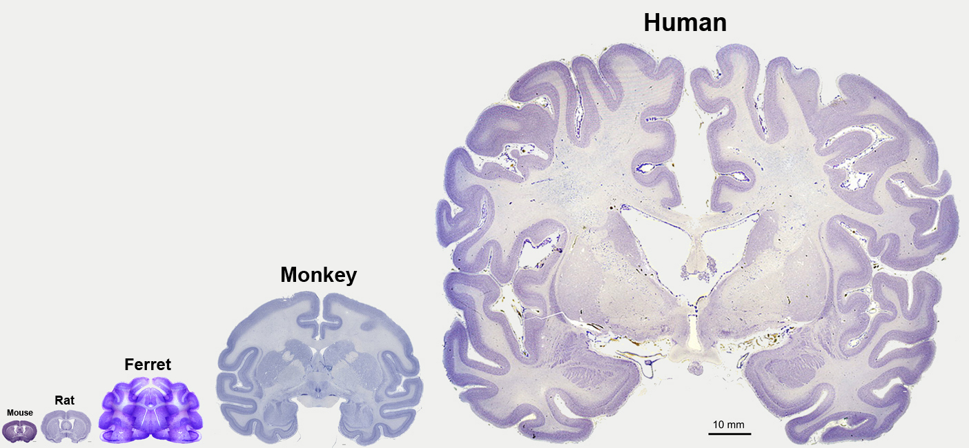
Some of these images were obtained from the MSU Human Brain Atlas & Brainmaps.org.